Metabolic and Immunologic Consequences of ABH Secretor Status |
|
PETER J. D'ADAMO and GREGORY S. KELLY Copyright 2000-2009 All Rights Reserved. Unauthorized reproduction prohibited by law.
This
monograph will attempt to organize and explore these associations in some
detail. Functional
and Genetic Factors Involved in ABH Secretion The
term "ABH secretor," as used in blood banking, refers to
secretion of ABO blood group antigens in fluids such as saliva, sweat,
tears, semen, and serum. If
people are ABH secretors, they will secrete antigens according to their
blood groups. For example, group O people will secrete H antigen, group A
people will secrete A and H antigens, etc.
Soluble (secreted) antigens are called substances. To test for
secretor status, an inhibition or neutralization test is done using
saliva. The principle of the
test is that if ABH antigens are present in a soluble form in a fluid
(e.g., saliva) they will neutralize their corresponding antibodies and the
antibodies will no longer be able to agglutinate red cells possessing the
same antigens. One
of the primary differences in physiology between secretors and
non-secretors has to do with qualitative and quantitative differences in
components of their saliva, mucus, and other body secretions.
ABH secretion is controlled by two alleles,
Se and se. Se
is dominant and se is recessive (or amorphic).
Approximately 80% of people are secretors (SeSe or Sese). In
the most rudimentary sense, the secretor gene (FUT2 at 19q13.3) codes for
the activity of the glycosyltransferases needed to assemble aspects of
both the ABO and Lewis blood
groups. This it does in concert with the gene for group O, or H (FUT1).
These enzymes are then active in places like goblet and mucous
gland cells, resulting in the presence of the corresponding antigens in
body fluids.(1) The
H antigens are indirect gene products expressed as fucose-containing
glycan units, residing on glycoproteins or glycolipids of erythrocyte
membranes or on mucin glycoproteins in secretions and are the fucosylated
glycans substrates for glycosyltransferases that give rise to the epitopes
for the A , B and Lewis blood group antigens.
The major difference between the two genes is in their pattern of
expression: the FUT1 (H) gene is expressed predominantly in erythroid
tissues giving rise to FUT1 (H enzyme) whose products reside on
erythocytes, whereas the FUT2 (Secretor) gene is expressed predominantly
in secretory tissues giving rise to FUT2 (Secretor enzyme) and to products
that reside on mucins in secretions.
When
alleles of both genes fail to express active enzymes, individuals bearing
them, in homozygous state, lack the substrates for the A or B
glycosyltransferases and do not express the A and B epitopes. Relationship
of ABH Secretor Status and Lewis System Since
FUT1 provides the glycans necessary for glycosyltransferases conversion
into the Lewis antigen in addition to ABH, the Lewis blood group
determinants are structurally related to determinants of the ABO and the
H/h blood group systems and the outcome of Lewis typing can also often be
used for the de facto determination of ABH secretor status. In the
presence of FUT2 alleles that express type 1 H determinants, the phenotype
will be Le (a-b+) but individuals in whom the FUT2 gene is not expressed
will be (Le a+b-). ABH
secretors are almost always Lewis (a-b+) since they convert all their
Lewis (a) antigen into Lewis (b). ABH non-secretors are always (Lewis a+b-)
since they lack the FUT2 dependent glycosyltransferase to accomplish this.
A small section (1-4% of the
population dependent on race) will be Lewis Double Negative (LDN; Lewis
(a-b-)) and for which Lewis
typing cannot be used to determine ABH secretor status. In these
individuals determination via saliva is necessary. However, it may be
helpful to think of LDN individuals as a special category of non-secretor,
since they do lack the Lewis b antigen (like the traditional ABH
non-secretors). In
most instances LDNs share the same metabolic consequences as ABH
non-secretors, and in a few, such as cardiovascular disease and insulin
resistance, actually have the most severe variations.
Table
1: Lewis blood types and their relationship to ABH secretors/non-secretor
status. Although
ABH secretor status is often thought of as an all or none situation, this
is generally not the case. In some ABH non-secretors (known as partial or
weak secretors) there will often be some form of active A or B blood group
substance in the saliva; however, the quantity and quality of these
substances is greatly reduced, predisposing them to similar functional
problems as other non-secretors. (3,4) Antigenic
Structures in Fluid Secretions There
are several advantages in having large quantities of blood type antigens
(both ABO and Lewis) secreted into saliva. First, salivary carbohydrate
structures found in mucins can aggregate some oral bacteria and also
constituents of pellicle and plaque. Since saliva of secretors contains
substantially more diversity and total carbohydrate than non-secretor
mucins, this places secretors at a bit of an advantage. Second, these same
blood type carbohydrate structures, because of the noted sweet tooth of
many dietary lectins, might actually place secretors at a significant
advantage with respect to binding some blood type specific dietary
lectins. In
the gastric mucosa of healthy individuals, the normal mucosa of secretors
is characterized by a uniform distribution of blood type antigens in the
pits. Healthy mucosa of non-secretors shows little staining for these
blood type antigens, but, instead, demonstrates significant quantities of
the I(Ma) antigen. This tendency to express the I (Ma) antigen will
subsequently have an impact on antibody capabilities, as we will see when
we discuss immunity. (5) PHYSIOLOGIC
MANIFESTATIONS Brush
Border Hydrolases ABO
blood group determines much of the enzyme activity in the tissue
(brush-border) of the intestine. At least six intestinal hydrolases have
ABO blood group antigenic determinants that are directly related to ABO
blood group. Basically, the intestinal glycoproteins of blood group A and
B individuals express A or B antigens, while blood group O subjects
express the H determinant. The expression of these ABH antigens is under
the control of the secretor gene; so these ABH antigens are not detected
in the hydrolases of non-secretor subjects. (6) ABH secretors have greater
quantities of free ABH antigens in the makeup of their intestinal
secretions; this has significant effects on bacterial and lectin adherence
to the gut microvilli. Intestinal
Alkaline Phosphatase Activity The
activity of intestinal alkaline phosphatase and serum alkaline phosphatase
is strongly correlated with ABH secretor phenotypes.
Independent of ABO blood group, ABH non-secretors have lower
alkaline phosphatase activity than ABH secretors. It has been estimated
that the serum alkaline phosphatase activity of non-secretors is only
about 20% of the activity in the secretor groups. (7-10)
The
intestinal component of alkaline phosphatase is involved with both the
breakdown of dietary cholesterol and the absorption of calcium. The
differences in intestinal alkaline phosphatase are almost exclusively
related to one fraction of the intestinal alkaline phosphatase. Normal
molecular mass intestinal alkaline phosphatase (NIAP) is present in the
serum of both secretors and non-secretors regardless of ABO blood group.
However, the high molecular mass intestinal alkaline phosphatase only
appears in serum of Lewis (a-b+) blood group secretors. (11) It
should be mentioned that in addition to ABH secretor status, ABO
polymorphism is also linked to the levels and persistence of intestinal
alkaline phosphatase. (88) Numerous studies have associated group O
individuals with the highest alkaline phosphatase activity and group A the
least. (89) These
findings suggest that the link between group O individuals and adaptation
to cholesterol-containing foods in the diet (such as meats) reaches its
greatest accommodation in group O secretors. Conversely, group A
non-secretors would have the lowest levels of intestinal alkaline
phosphatase and the greatest difficulties in handling dietary fat. In
addition, one study has implied that the group A antigen itself may
inactivate IAP. (90) Bacterial
Flora The
role of ABO blood group in
determining some of the bacteria making up a healthy GI ecosystem is
particularly strong in ABH secretors. Since ABH secretor status and ABO
blood group dictate the presence and specificity of A, B, and H blood
group antigens in human gut mucin glycoproteins, this can influence the
populations of bacteria capable of taking up local residence. This occurs
because some of the bacteria in the digestive tract are actually capable
of producing enzymes that allow them to degrade the terminal sugar of the
ABH blood type antigens for a constant food supply. For
example, bacteria capable of degrading blood group B antigen produce
enzymes that that allow them to detach the terminal alpha-D-galactose and
use this sugar for food. Blood
group A degrading bacteria would have similar capabilities with respect to
N-acetylgalactosamine. Group B secretors produce greater levels of
B-degrading than A- or H-degrading activity, and A secretors produce
greater levels of A-degrading than B- or H-degrading activity. Because of
this capability, the bacteria that use ABH antigens for food have a
competitive advantage and can thrive in the environment created by the
preconditioning of ABH secretions. Although
comparatively small populations of bacteria produce blood group-degrading
enzymes (estimated populations are 10(8) per g); the quantity of these
bacteria are several orders of magnitude greater in different blood types
and are much more stable residents. For example, B-degrading bacteria have
a population density of about 50,000-fold greater in blood group B
secretors than in other subjects. Similar bacterial specificity and enzyme
activity is found among other blood types. (12,13) Breast
Milk Components Significant
variations in the carbohydrate residues in human breast milk are found
depending on the mothers ABO, Lewis, and Secretor blood types. During the
first week of lactation the ability to produce neuraminyloligosaccharides
is linked to the ABH secretor groups. And the ability to produce
oligosaccharides with Le (a) or Le (b) characteristics is linked to Lewis
and Secretor systems. The consequences of this are that secretors will
produce higher levels of N-acetylneuraminic acid and lower levels of
galactose in their breast milk than non-secretors. In the ABH secretor
groups, blood type A and O secretors also have higher N-acetylglucosamine
contents than B and AB secretors (p less than 0.001), while the A and B
secretors have higher galactose levels. The Lewis secretor groups are also
distinguished by a significantly higher level of fucose. The ABH (+), Le
(a-b-) group had higher lactose contents than the other groups. (28)
Table
2. Differences in carbohydrate composition by ABO and Lewis blood groups
and ABH secretor status.
Blood
Clotting ABO
blood group impacts the clotting ability to a significant degree,
in fact, it has been estimated that a significant fraction (30%) of the
genetically determined variance in plasma concentration of the von
Willebrand factor antigen (vWf) is directly related to ABH determinants.
As a rule, it is blood group O individuals who have the lowest amount of
this clotting factor. (29) ABH
non-secretors are reported to have shorter bleeding times and a tendency
towards higher factor VIII Among
persons belonging to blood group O (the blood type most likely to have
problems with clotting), the lowest concentration of vWf:Ag and VIII:Ag is
found in the group O secretors. While blood group O non-secretors will
have a higher concentration of both vWf:Ag and factor VIII antigen (VIII:Ag),
providing them with a better capability for clotting. (32) Among
blood groups A, B, and AB, also having the Lewis (a- b-) phenotype is
associated with the highest degree of clotting factors. In white men with
these blood types, the Lewis (a-b-) phenotype, infers significantly higher
levels of factor VIII and von Willebrand factor. Among black men with
blood type A, B, or AB, and phenotype Lewis (a-b-), a similar trend is
found with these individuals having the highest values for factor VII and
von Willebrand factor. In women with blood type A, B, or AB, and phenotype
Le(a-b-) a correlation exists for higher levels of factor VIII.
Table
3: Lewis Blood Type and Clotting Factors
Based
upon this research, researchers have suggested that the Le (a-b-)
phenotype (and blood groups A, B, and AB especially), by virtue of their
association with raised levels of factor VIII and von Willebrand factor,
might be at a higher risk for future thrombotic and heart disease. (33) Dental
Cavities In
all blood groups, the average amount of cavities is lower for ABH
secretors than for non-secretors. This difference is most significant for
smooth surface areas of the teeth. Also, secretors of blood group A had
the lowest numbers of cavities. (34) Diabetes,
Heart Disease, & Metabolic Syndrome X Diabetes ABH non
secretors, and especially Lewis negative individuals, are at a greater
risk of developing diabetes (especially adult onset diabetes); and they
might be at a greater risk of developing complications from diabetes.
Findings suggest that an increased proportion of non-secretors are found
among patients with diabetes, particularly of the insulin-dependent
diabetes type. (14,15) The Lewis
negative (Le a-b-) red blood cell phenotype appears to confer the greatest
risk of developing diabetes. This
blood type is observed more than three times more frequently (29%) in
diabetics irrespective of their clinical type.
Non-diabetics categorized as low insulin responders to glucose are
also significantly more likely to be Lewis negative.
(16) Among individuals
with juvenile diabetes mellitus, the prevalence of severe retinopathy (a
side effect of diabetes) is lower in ABH secretors than in the ABH
non-secretor group. (17) Heart
Disease Data allows the
conclusion that the ABH non-secretor phenotypes are a risk factor for
myocardial infarction, this is particularly true for recessive Lewis blood
types and even more so among men than women. ABH secretors seem to have
been given a bit of genetic resistance against heart disease while Lewis
negative individuals appear to be at the highest risk for CHD.
This finding was
reported in the Copenhagen Male study and replicated in the NHLBI Family
Heart Study. Eight percent of men with the Lewis (a-b-) phenotype had a
history of non-fatal myocardial infarction (among Lewis positive men the
frequency was only 4%). But
even worse, research showed that men with Lewis (a-b-) had an increased
risk of death from ischemic heart disease (IHD) (IHD case fatality rate
(RR = 2.8 (1.5-5.2), P = 0.01)) compared with others.
Adjusted for age, relative risk climbed even higher to 4.4
(1.9-10.3), P < 0.001, and for all causes of mortality RR = 1.6
(1.0-2.6), P < 0.05. (18) Results from the
NHLBI Family Heart Study also showed a higher risk of coronary heart
disease (odds ratio was 2.0 (95% confidence interval = 1.2 to 3.1) for
Lewis (a-b-) versus other Lewis groups.
Triglycerides were significantly higher in the Lewis
(a-b-) subjects. Among
women, there was also a trend towards increased risk of CHD among Lewis
negative phenotypes; however, the trend is dramatically weaker than among
male subjects. (19) Additional
research has also duplicated these results, supporting and adding to the
weight of evidence linking Lewis negative phenotype Lewis (a-b-) as a
marker of high risk for the development of ischemic heart disease.
Even excluding the Lewis negative phenotype, the secretor phenotype
Lewis (a-b+) was found to be a genetic marker of resistance against the
development of ischemic heart disease, while ABH non-secretor status is a
risk factor predisposing individuals towards heart disease. (20) Protective
Effects of Alcohol In men Lewis
(a-b-), the Lewis negative or excessive phenotype, a group genetically at
high risk of ischemic heart disease (IHD), alcohol consumption seems to be
especially protective. In the Copenhagen study, researchers found that
drinking alcohol was the only risk factor that had an interaction with
Lewis negative blood type and that alcohol could strongly modify risk in
an inverse (so hence positive) manner.
There was a significant inverse dose-effect relationship between
alcohol consumption and decreasing risk. (21) Alcoholism
and Alcohol's Protective Benefits Paradoxical with
the cardiovascular benefits of alcohol in Lewis negative individuals,
several large studies have associated alcoholism with ABH non-secretor
status. (22,23) Metabolic
Syndrome X Data suggest that
Lewis (a-b-) men exhibit features of the insulin resistance syndrome or
syndrome X, including a tendency to prothrombic metabolism and higher
levels of BMI, SBP, triglycerides, and fasting levels of serum insulin and
plasma glucose. These same relationships are not as strong for women. A group of
metabolic problems comprised of insulin resistance, elevated plasma, lipid
regulation problems (elevated triglycerides, increased small low-density
lipoproteins, and decreased high-density lipoproteins), high blood
pressure, a prothrombic state, and obesity (especially central obesity or
a predisposition to gaining weight in the abdomen) combine to form
"Metabolic Syndrome X" (MSX).
This cluster of metabolic disorders seems
to promote the development of diabetes (adult onset type II),
arteriosclerosis, and cardiovascular disease.
And while insulin resistance might lie at the heart of the problem,
all of these metabolic disorders appear to contribute to health problems. Because of the
associations with non-secretor status and both diabetes and heart disease,
many different researchers have explored the connection between a
metabolic syndrome called "Syndrome X" and Lewis and
non-secretor blood types. Just as is the case with diabetes and heart disease,
individuals with Lewis (a-b-) phenotype are most predisposed to MSX.
It has even been hypothesized that Lewis
(a-b-) men and syndrome X share a close genetic relationship on
chromosome 19 and that the Lewis (a-b-) phenotype is a genetic marker of
the insulin resistance syndrome. (24) As we will
discuss in the next section on clotting, non-secretors and especially
Lewis negative individuals, are especially prone to prothrombic metabolism
(a tendency to form clots more readily and to have slower bleeding times).
The tendency to higher triglycerides was mentioned when we
discussed heart disease. (25) Researchers have
also investigated Lewis blood types as part of the Copenhagen Study, and
they found very supportive evidence of trends toward metabolic
differences. Compared to all
other men, the Le (a-b-) men had a significantly higher systolic blood
pressure (6 mm Hg, P = .0024). They also had higher values of body mass
index (8%, P = .016), total body fat mass (25%, P = .015), fasting values
of serum insulin (32%, P = .006), serum C-peptide (20%, P = .029), and
plasma glucose (8%, P = .003). These trends, while consistent for men,
were not as strong for women. (26) IMMUNOLOGIC
CONSEQUENCES Basic
Functions Evidence
suggests that ABH non-secretors have lower levels of IgG. (35,36) In tests of 202 Caucasians researchers found IgA concentrations to be significantly lower in
non-secretors than in secretors. (37,38) This seems to imply that the ABH
non-secretor state is associated with a "Defense In Depth"
strategy (i.e. let the invader in and attempt to destroy it internally)
versus the ABH secretor state, which implies a "Preclusive
Strategy" (i.e. wall out the invader and don't allow entrance in the
first place.) For example, the free ABH antigen on the mucosa barriers of
ABH secretors acts as an effect anti-adhesive mechanism against ABH
specific bacterial fimbrae lectins. On
the other hand, the ability to secrete relatively different concentration
of the components of the blood group substances as determined by
secretors/non-secretor genetics seems to affect phagocytic activity of the
leucocytes in a manner that actually places non-secretors at somewhat of
an advantage. In general, leukocytes of non-secretors have substantially
greater ingestion power as compared to secretors. Although this ability
appears to be across the board for all non-secretors, blood group
O and B non-secretors have the greatest advantage and highest
phagocytic activity. (41) Perhaps
this is a compensatory mechanism for their more limited antigenic barrier
in their body fluids and secretions. Results
suggest that the level of anti-I in the serum of normal individuals may be
affected by the donor's ABO group, secretor status and sex. For
individuals with blood group O, B and AB secretors have higher levels of
an antibody presumed to be auto-anti-I (cold hemagglutinin). The level of
this antibody is usually even higher among non-A female secretors than for
males. (39) Researchers
have found that in individuals with insulin dependent diabetes mellitus,
the mean level of C3c for non-secretors is significantly lower than that
found for secretors. The
level of C4 among ABH non-secretors
was also significantly lower than that of ABH secretors. (40) Helicobacter pylori The
genetics of the ABH secretor/non-secretor system interact to alter an
individual's risk for ulcers. In several studies, non-secretors of ABH
substances have been found to have a significantly higher rate of duodenal
and peptic ulcers. (42,43) In
fact the Copenhagen study found that the lifetime prevalence of peptic
ulcer in men who were ABH non-secretors
was 15% (statistically 15% of ABH non-secretors will have an ulcer at some
point in their lives). And, the attributable risk of peptic ulcer in men
who were Lewis (a + b-) or ABH non-secretors, with blood group O or A
phenotypes was 37%. (44) Overall,
the relative risk of gastroduodenal disease for non-secretors compared
with secretors is 1.9 (95% confidence interval). Duodenal ulcer patients
are more likely to be non-secretors, and being a non-secretor acts as a
multiplicative risk factor with the gene for hyperpepsinogenemia I to
impact the risk of duodenal ulcer. (45,46) Because
of the increased prevalence of ulcers among non-secretors researchers have
suggested that secretor status might influence bacterial colonization
density or the ability of H. pylori to attach to gastroduodenal cells.
With regards to the overall interaction with H. pylori infection,
non-secretor status is generally considered to be a separate independent
risk factor for gastroduodenal disease in addition to H. pylori infection;
however, there is more to this story, and, in fact some interesting
interactions between secretor status, Lewis genetics, and H. pylori.
(47) Because
non-secretors are limited in their ability to secrete the Lewis (b) blood
group antigen into the mucus secretions of their digestive tract, it has
been proposed that they be at a competitive disadvantage from preventing
H. pylori attachment. In fact, the Lewis (b) antigens have been found to
act as somewhat of a preferential target for H. pylori attachment.
Thus, lack of Lewis (b) in mucosal fluids of ABH non secretors
might indirectly contribute to colonization by H. pylori. (48-50) In
a simplified sense, when the Lewis (b) antigen is free floating in the
mucus, it probably acts to bind up some of the H. pylori before it can
contact and attach to host tissue. In essence, being an ABH secretor
provides an ability to put some biological decoys or metabolic chaff out
into the gastric secretions that is very specific for H. pylori. Also, in
ABH non-secretors the
immune response against H. pylori appears to be lower and H. pylori
appears to attach with higher aggressiveness and cause more inflammation.
(51) Individuals
with Lewis (a+b-) ABH non-secretor phenotype also show a significantly
higher proportion of the H. pylori-seronegative subjects and a lower IgG
(H. pylori immunoglobulin G (IgG) antibody) immune response to H. pylori
antigens as compared with the individuals of Lewis (a-b+)/secretor
phenotype. Evidence
also indicates that 100% of non-secretors with duodenal ulcers culture
positive for H. pylori infection. However, among non-secretors with
gastric ulcer, H. pylori is found in only about 12.5% of the cases. This
is not observed among secretors, who are nearly equally likely to have H.
pylori infection in either gastric or duodenal ulcer.
(52) Bacteria
Urinary Tract Infections ABH
non-secretors are at a greater risk for recurrent urinary tract infections
(UTI) and are much more likely to develop renal scars. This susceptibility
is even greater among the Lewis negative subset (Le (a-b-)).
The ABH secretor phenotype conveys
a measure of protection; cutting the risk of recurrent UTI
by greater than 50% and dramatically decreasing the likelihood that
renal scars will develop. ABH
non-secretors appear to be at extra risk for recurrent urinary tract
infections. In one study of women with recurrent UTI, 29 % of the women
were the Lewis (a+ b-) non-secretor phenotype, while another 26% of the
women were Lewis (a- b-) recessive phenotype. When the women with ABH
non-secretor and recessive phenotypes were combined and considered
collectively, the odds ratio (an estimate of relative risk of recurrent
urinary tract infection) for those without the secretor phenotype (Lewis
(a-b+) was 3. (53-57) A
form of synergy also appears to exist between UTI risk, secretor status
and the lack of ability to create anti-B isohemagglutinin. Essentially,
blood group B and AB and the
non-secretor phenotype seem to work together to increase the relative risk
of recurrent UTI among these women. (58)
Evidence also indicates that women and children with renal scarring
subsequent to recurrent UTI and pyelonephritis are more likely to be ABH
non-secretors. (59-61) As many as 55-60% of all ABH non-secretors have
been found to develop renal scars, even with the regular use of antibiotic
treatment for UTI whereas as
few as 16% of ABH secretors will develop similar renal scarring. (62) This
tendency to scarring does not seem to be dictated as much by the
aggressiveness of the bacterial infection, but by the more aggressive
inflammatory response created by ABH non-secretors against the bacterial
infection. The levels of C-reactive protein, erythrocyte sedimentation
rate and body temperature are significantly higher in non-secretors than
in secretors (p less than 0.04) with recurrent UTI. As a consequence,
non-secretors seem to self inflict to a degree the renal scarring
secondary to their acute phase inflammatory response. (63) Neisseria
sp. The
genetically determined inability to secrete the water-soluble glycoprotein
form of the ABO blood group antigens into saliva and other body fluids is
a recognized risk factor for Neisseria meningococcal disease. ABH non-secretors are consistently over represented among individuals
contracting this infection. This over representation is even
greater among individuals who are carriers of the infection. (64) Secretory immune capabilities and other factors appear
to contribute to the relative protection against colonization by
meningococci enjoyed by ABH secretors.
ABH non-secretors
typically have lower levels of anti-meningococcal salivary IgM, and if to
add insult to injury, both the IgA and IgM antibodies produced by ABH
secretors are more effective at providing protection against this
microorganism. (65) Candida
sp. ABH
non-secretors are much more likely to be carriers of Candida sp. and to
have problems with persistent Candida infections. Blood group O
non-secretors are the most affected of the non-secretor blood types. One
of the innate defenses against superficial infections by Candida species
appears to be the ability of an individual to secrete the water-soluble
form of his ABO blood group antigens into body fluids. The protective
effect afforded by the secretor gene might be due to the ability of
glycocompounds in the body fluids of secretors to inhibit adhesins
(attachment lectins) on the surface of the yeast. In attachment studies,
preincubation of blastospores with boiled secretor saliva significantly
reduced their ability to bind to epithelial cells. ABH non secretor saliva
did not reduce the binding and often enhanced the numbers of attached
yeasts. (66,67) In one study, among individuals with Type II diabetes, 44%
of ABH non-secretors were
oral carriers of this yeast. (68) Although
non-secretors make up only about 26% of the population, they are
significantly over represented among individuals with either oral or
vaginal Candida infections, making up almost 50% of affected individuals.
(69) The inability to secrete blood group antigens in saliva also
appears to be a risk factor in the development of, or persistence of
chronic hyperplastic Candidosis. In one study, the proportion of
non-secretors of blood group antigens among
patients with chronic hyperplastic Candidosis was 68%.
(70) Women
with recurrent idiopathic vulvovaginal Candidiasis are much more likely to
be ABH non-secretors.
Combining both ABH non-secretor phenotype and absence of the Lewis gene
Lewis (a- b-), the relative risk of chronic recurring vulvovaginal
Candidiasis is between 2.41-4.39, depending on the analysis technique and
control group. (71) Oral
carriage of Candida is also significantly associated with blood group O (p
less than 0.001) and independently, with non-secretion of blood group
antigens (p less than 0.001), with the trend towards carriage being
greatest in group O non-secretors. (72) Autoimmune
Disease ABH
non-secretors appear to have an increase in the prevalence of a variety of
autoimmune diseases including ankylosing spondylitis, reactive arthritis,
psoriatic arthropathy, Sjogren's syndrome, multiple sclerosis, and Grave's
disease. This susceptibility towards autoimmune problems appears to be
most pronounced among Lewis (a-b-) phenotypes.
Among individuals with spondyloarthropathies, non-secretors are
reported to make up 47% of the patient population. In the subgroup of
these patients suffering from ankylosing spondylitis, ABH
non-secretors account for 49% of patients. Since the control population had a prevalence of
non-secretors of 27% (consistent with the expected percent in the general
population), it appears that in spondyloarthropathies in general, and
ankylosing spondylitis specifically, non-secretors are dramatically over
represented. (73,74) Among
individuals with primary Sjogren's syndrome, Lewis blood group frequency
differs from that of the general population, due mainly to an increased
Lewis negative phenotype (Le (a-b-)) frequency. (75) The
inability to secrete the water soluble glycoprotein form of the ABO blood
group antigens into saliva is significantly more common in patients with
Graves' disease than control subjects (40% versus 27%: p less than 0.025)
but not among those with Hashimoto's thyroiditis or spontaneous primary
atrophic hypothyroidism. ABH
non-secretors with Grave's disease were found to produce higher levels of
antitubulin antibodies, while levels of other antibodies were similar to
secretors. (76) Celiac
Disease ABH
Non-secretors are at an increased risk for development of celiac disease.
One study reported that to
48% of patients with celiac disease were reported to be ABH non-secretors.
(77) This appears to be especially true for the recessive Lewis (a-b-)
phenotype. Evidence suggests an increased prevalence of complications and
celiac-associated abnormalities is also found in the non-secreting and
negative Lewis celiac patients. (78) Pulmonary
Considerations ABH
secretors are significantly over represented among patients with influenza
viruses A and B (55/64, 86%; p less than 0.025), rhinoviruses (63/72, 88%;
p less than 0.01), respiratory syncytial virus (97/109, 89%; p less than
0.0005), and echoviruses (44/44, p less than 0.0005). Why this increased
risk appears in secretors has not been clearly established. (79)
Among
coal miners, asthma was significantly related to non-secretor phenotype.
In this population, significantly lower lung function and higher
likelihood of wheezing is especially prevalent among Lewis-negative or
non-secretor subjects with blood group O. (80) Independent findings
suggest that the ability to secrete ABH antigens might decrease the risk
of COPD. Non-secretors have been found to have significantly greater
impairment of forced expiration. ABH non secretors have lower mean values
of forced expiratory volume in one second as a percentage of forced vital
capacity (FEV1/FVC%) and a significantly larger proportion of them had
aberrant values, defined as FEV1/FVC% less than 68.
(81) ABH
non-secretor status also offers a slight increase risk for habitual
snoring. (82) NEOPLASIA
AND MALIGNANCY Secretor
and Lewis Phenotypes and Tumor Markers Accurately
predicting the relevance of some tumor markers for diagnosis of cancer
appears to be dependent on both secretor status and Lewis blood group. As
an example, some researchers have suggested that taking into account
aspects of Lewis and/or Secretor status in order to establish reference
ranges might actually be a way to increase the clinical utility of the CA
19-9 tumor marker. (83) There
is a substantial difference in levels of this tumor marker are under the
control of Secretor and Lewis genetics. Individuals having homozygous
inactive Se alleles (se/se) and homozygous active Le alleles
(Le/Le), exhibited the highest mean CA19-9 value. All of the Lewis
negative individuals (Le (a- b-) consisting of a le/le genotype) had
completely negative CA19-9 values, irrespective of the Se genotype.
On
the other hand, Lewis negative individuals showed a higher mean DU-PAN-2
value than did the Le-positive individuals. Among patients with colorectal
cancer, the Le-negative patients (le/le) with colorectal cancer showed
undetectable CA19-9 values, i.e., less than 1.0 unit/ml, but many of them
exhibited highly positive DU-PAN-2 values. In contrast, many of the
Le-positive patients (Le/Le or Le/le) had positive CA19-9 values, whereas
very few of them exhibited positive DU-PAN-2 values. (84)
Table
4: CA19-9 and DU-PAN-9 expression in
colorectal cancer correlated to Lewis subtype. This implies that the CA19-9 measurement is not a useful tumor marker for Le-negative individuals, although DU-PAN-9 appears to be. Lewis negative individuals do not express any kinds of type 1 chain Lewis antigens (Lewis (a), Lewis (b), and secretory Lewis(a)) in their digestive organs. It is, therefore, not useful to measure the CA19-9 titer of the Lewis negative cancer patient. (85) Preneoplastic
Changes and Cancer As
a general rule, a higher intensity of oral disease is found among ABH
non-secretors. So it is not surprising that when it comes to precancerous,
or cancerous changes to tissue of the mouth and esophagus, ABH
non-secretors seem to fair worse than ABH secretors. This oral disease
susceptibility is reflected in the occurrence of epithelial dysplasia, for
example, which is found almost exclusively in the non-secretor group. (86) Barrett's
esophagus, a condition often preceding the development of esophageal
cancer, and esophageal cancer also have a positive association with Lewis
(a+b-) non-secretor phenotypes. (87) ACKNOWLEDGEMENTS The
author wishes to thank Gregory Kelly, ND for his help in the preparation
of this manuscript.
ABH SECRETOR STATUS TESTING A single saliva-based test can be ordered from North American Pharmacal. CONTACT
ADDRESS 2009
Summer Street http://www.dadamo.com REFERENCES 1. Bals R, Woeckel W, Welsch U. Use of antibodies directed against blood group substances and lectins together with glycosidase digestion to study the composition and cellular distribution of glycoproteins in the large human airways. J Anat 1997 Jan;190 ( Pt 1):73-84 2.
Prakobphol A, Leffler H,
Fisher SJ. The high-molecular-weight human mucin is the primary salivary
carrier of ABH, Le(a), and Le(b) blood group antigens. Crit Rev Oral Biol
Med 1993;4(3-4):325-33 3.
Mohn JF, Owens NA,
Plunkett RW. The inhibitory properties of group A and B non-secretor
saliva. Immunol Commun 1981;10(2):101-26 4.
Kogure T, Furukawa K.
Enzymatic conversion of human group O red cells into Group B active cells
by alpha-D-galactosyltransferases of sera and salivas from group B and its
variant types. J Immunogenet 1976 Jun;3(3):147-54 5.
Kapadia A, Feizi T,
Jewell D, et al. Immunocytochemical studies of blood group A, H, I, and i
antigens in gastric mucosae of infants with normal gastric histology and
of patients with gastric carcinoma and chronic benign peptic ulceration. J
Clin Pathol 1981 Mar;34(3):320-37 6.
Triadou N, Audran E,
Rousset M, et al. Relationship between the secretor status and the
expression of ABH blood group antigenic determinants in human intestinal
brush-border membrane hydrolases. Biochim Biophys Acta 1983 Dec
27;761(3):231-6 7.
Domar U, Hirano K,
Stigbrand T. Serum levels of human alkaline phosphatase isozymes in
relation to blood groups. Clin Chim Acta 1991 Dec 16;203(2-3):305-13 8.
Mehta NJ, Rege DV,
Kulkarni MB. Total serum alkaline phosphatase (SAP) and serum cholesterol
in relation to secretor status and blood groups in myocardial infarction
patients. Indian Heart J 1989 Mar;41(2):82-85 9.
Tibi L, Collier A,
Patrick AW, et al. Plasma alkaline phosphatase isoenzymes in diabetes
mellitus. Clin Chim Acta 1988 Oct 14;177(2):147-155 10.
Agbedana EO, Yeldu MH.
Serum total, heat and urea stable alkaline phosphatase activities in
relation to ABO blood groups and secretor phenotypes. Afr J Med Med Sci
1996 Dec;25(4):327-9 11.
Matsushita M, Irino T,
Stigbrand T, et al. Changes in intestinal alkaline phosphatase isoforms in
healthy subjects bearing the blood group secretor and non-secretor. Clin
Chim Acta 1998 Sep 14;277(1):13-24 12.
Hoskins LC, Boulding ET.
Degradation of blood group antigens in human colon ecosystems. II. A gene
interaction in man that affects the fecal population density of certain
enteric bacteria. J Clin Invest 1976 Jan;57(1):74-82 13.
Hoskins LC, Boulding ET.
Degradation of blood group antigens in human colon ecosystems. I. In vitro
production of ABH blood group-degrading enzymes by enteric bacteria. J
Clin Invest 1976 Jan;57(1):63-73
14.
Patrick AW, Collier A. An
infectious aetiology of insulin-dependent diabetes mellitus?
Role of the secretor status. FEMS
Microbiol Immunol 1989 Jun;1(6-7):411-416 15.
Peters WH, Gohler W. ABH-secretion and Lewis red cell groups in diabetic
and normal subjects from Ethiopia. Exp
Clin Endocrinol 1986 Nov;88(1):64-70 16.
Melis C, Mercier P, Vague P, Vialettes B. Lewis antigen and diabetes.
Rev Fr Transfus Immunohematol 1978 Sep;21(4):965-71 17.
Eff C, Faber O, Deckert T. Persistent
insulin secretion, assessed by plasma C-peptide estimation in long-term
juvenile diabetics with a low insulin requirement.
Diabetologia 1978 Sep;15(3):169-72 18.
Hein HO, Sorensen H, Suadicani P, Gyntelberg F.
The Lewis blood group--a new genetic marker of ischaemic heart
disease. J Intern Med 1992
Dec;232(6):481-7 19.
Ellison RC, Zhang Y, Myers RH, et al.
Lewis blood group phenotype as an independent risk factor for
coronary heart disease (the NHLBI Family Heart Study).
Am J Cardiol 1999 Feb 1;83(3):345-8 20.
.Zhiburt BB, Chepel' AI, Serebrianaia NB, The Lewis antigen system as a
marker of IHD risk. Ter Arkh
1997;69(1):29-31 [Article in Russian] Slavchev S, Tsoneva M, Zakhariev Z.
The secretory type of persons who have survived a myocardial
infarct. Vutr Boles
1989;28(2):31-4 [Article in Bulgarian] 21.
Hein HO, Sorensen H, Suadicani P, Gyntelberg F.
Alcohol intake, Lewis phenotypes and risk of ischemic heart
disease. The Copenhagen Male
Study. Ugeskr Laeger 1994 Feb
28;156(9):1297-302 22.
Cruz-Coke R. Genetics and
alcoholism. Neurobehav
Toxicol Teratol 1983 Mar-Apr;5(2):179-80 23.
Kojic T, Dojcinova A, Dojcinov D, et al. Possible genetic predisposition for alcohol addiction.
Adv Exp Med Biol 1977;85A:7-24 24.
Petit JM, Morvan Y,
Viviani V, Vaillant G, Matejka G, Rohmer JF, Guignier F, Verges B, Brun JM. Related Articles Insulin resistance syndrome and Lewis
phenotype in healthy men and women. Horm
Metab Res. 1997
Apr;29(4):193-5 25.
Hein HO, Sorensen H, Suadicani P, Gyntelberg F.
Alcohol consumption, Lewis phenotypes, and risk of ischaemic heart
disease. Lancet 1993 Feb
13;341(8842):392-6 26.
Petit JM, Morvan Y, Mansuy-Collignon S, Viviani V, et al.
Hypertriglyceridaemia and Lewis (A-B-) phenotype in
non-insulin-dependent diabetic patients. Diabetes Metab 1997 Jun;23(3):202-4 27.
Clausen JO, Hein HO, Suadicani P, et al. Lewis phenotypes and the insulin resistance syndrome in young
healthy white men and women. Am
J Hypertens 1995 Nov;8(11):1060-6
28.
Viverge D, Grimmonprez
L, Cassanas G, et al. Discriminant carbohydrate components of human milk
according to donor secretor types. J Pediatr Gastroenterol Nutr 1990
Oct;11(3):365-70 29.
Orstavik KH, Kornstad L,
Reisner H, Berg K. Possible effect of secretor locus on plasma
concentration of factor VIII and von Willebrand factor. Blood 1989
Mar;73(4):990-3 30.
Wahlberg TB, Blomback M,
Magnusson D. Influence of sex, blood group, secretor character, smoking
habits, acetylsalicylic acid, oral contraceptives, fasting and general
health state on blood coagulation variables in randomly selected young
adults. Haemostasis 1984;14(4):312-9 31.
Orstavik KH. Genetics of
plasma concentration of von Willebrand factor. Folia Haematol Int Mag Klin
Morphol Blutforsch 1990;117(4):527-31 32.
Orstavik KH, Kornstad L,
Reisner H, Berg K. Possible effect of secretor locus on plasma
concentration of factor VIII and von Willebrand factor. Blood 1989
Mar;73(4):990-3 33.
Green D, Jarrett O, Ruth
KJ, Folsom AR, Liu K. Relationship among Lewis phenotype, clotting
factors, and other cardiovascular risk factors in young adults. J Lab Clin
Med 1995 Mar;125(3):334-339 34.
Arneberg P, Kornstad L,
Nordbo H, Gjermo P. Less dental caries among secretors than among
non-secretors of blood group substance. Scand J Dent Res 1976
Nov;84(6):362-6 35.
Al-Agidi SK, Shukri SM.
Association between immunoglobulin levels and known genetic markers in an
Iraqi population. Ann Hum Biol 1982 Nov-Dec;9(6):565-9 36.
Shinebaum R. ABO blood
group and secretor status in the spondyloarthropathies. FEMS Microbiol
Immunol 1989 Jun;1(6-7):389-95 37.
Grundbacher FJ.
Immunoglobulins, secretor status, and the incidence of rheumatic
fever and rheumatic heart disease. Hum Hered. 1972;22(4):399-404. 38.
Grundbacher FJ. Genetic
aspects of selective immunoglobulin A deficiency.
J Med Genet. 1972 Sep;9(3):344-7. 39.
Dube VE, Tanaka M,
Chmiel J, Anderson B. Effect of ABO group, secretor status and sex on cold
hemagglutinins in normal adults. Vox Sang 1984;46(2):75-9 40.
Blackwell CC, Weir DM,
Patrick AW, Collier A, Clarke BF. Secretor state and complement levels (C3
and C4) in insulin dependent diabetes mellitus. Diabetes Res 1988
Nov;9(3):117-9 41.
Tandon OP, Bhatia S,
Tripathi RL, Sharma KN. Phagocytic response of leucocytes in secretors and
non-secretors of ABH (O) blood group substances. Indian J Physiol
Pharmacol 1979 Oct-Dec;23(4):321-4 42.
Odeigah PG. Influence of
blood group and secretor genes on susceptibility to duodenal ulcer. East
Afr Med J 1990 Jul;67(7):487-500 43.
Suadicani P, Hein HO,
Gyntelberg F. Genetic and life-style determinants of peptic ulcer. A study
of 3387 men aged 54 to 74 years: The Copenhagen Male Study. Scand J
Gastroenterol 1999 Jan;34(1):12-7 44. Hein HO, Suadicani P, Gyntelberg F. Genetic markers for stomach ulcer. A study of 3,387 men aged 54-74 years from The Copenhagen Male Study. Ugeskr Laeger 1998 Aug 24;160(35):5045-46 45.
Dickey W, Collins JS,
Watson RG, et al. Secretor status and Helicobacter pylori infection are
independent risk factors for gastroduodenal disease. Gut 1993
Mar;34(3):351-3 46.
Sumii K, Inbe A, Uemura
N, et al. Multiplicative effect of hyperpepsinogenemia I and non-secretor
status on the risk of duodenal ulcer in siblings. Gastroenterol Jpn 1990
Apr;25(2):157-61 47.
Dickey W, Collins JS,
Watson RG, et al. Secretor status and Helicobacter pylori infection are
independent risk factors for gastroduodenal disease. Gut 1993
Mar;34(3):351-3 48.
Oberhuber G, Kranz A,
Dejaco C, et al. Blood groups Lewis(b) and ABH expression in gastric
mucosa: lack of inter-relation with Helicobacter pylori colonisation and
occurrence of gastric MALT lymphoma. Gut 1997 Jul;41(1):37-42 49.
Su B, Hellstrom PM,
Rubio C, et al. Type I Helicobacter pylori shows Lewis(b)-independent
adherence to gastric cells requiring de novo protein synthesis in both
host and bacteria. J Infect Dis 1998 Nov;178(5):1379-90 50.
Alkout AM, Blackwell CC,
Weir DM, et al. Isolation of a cell surface component of Helicobacter
pylori that binds H type 2, Lewis(a), and Lewis(b) antigens.
Gastroenterology 1997 Apr;112(4):1179-87 51.
Klaamas K, Kurtenkov O,
Ellamaa M, Wadstrom T. The Helicobacter pylori seroprevalence in blood
donors related to Lewis (a,b) histo-blood group phenotype. Eur J
Gastroenterol Hepatol 1997 Apr;9(4):367-70 52.
Mentis A, Blackwell CC, Weir DM, et al. ABO blood group, secretor
status and detection of Helicobacter pylori among patients with gastric or
duodenal ulcers. Epidemiol Infect 1991 Apr;106(2):221-9 53.
Sheinfeld J, Schaeffer AJ, Cordon-Cardo C, et al. Association of
the Lewis blood-group phenotype with recurrent urinary tract infections in
women. N Engl J Med 1989 Mar 23;320(12):773-7 54.
Similar findings by other researchers support this over
representation of recurrent UTI among non-secretors both in women and
children. 55.
May SJ, Blackwell CC,
Brettle RP, MacCallum CJ, Weir DM. Non-secretion of ABO blood group
antigens: a host factor predisposing to recurrent urinary tract infections
and renal scarring. FEMS Microbiol Immunol 1989 Jun;1(6-7):383-7 56.
A, Nudelman E, Clausen
H, et al. Binding of uropathogenic Escherichia coli R45 to glycolipids
extracted from vaginal epithelial cells is dependent on histo-blood group
secretor status. J Clin Invest 1992 Sep;90(3):965-72 57.
Jantausch BA, Criss VR,
O'Donnell R, et al. Association of Lewis blood group phenotypes with
urinary tract infection in children. J Pediatr 1994 Jun;124(6):863-8 58.
Kinane DF, Blackwell CC,
Brettle RP, et al. ABO blood group, secretor state, and susceptibility to
recurrent urinary tract infection in women. Br Med J (Clin Res Ed) 1982
Jul 3;285(6334):7-9 59.
May SJ, Blackwell CC,
Brettle RP, MacCallum CJ, Weir DM. Non-secretion of ABO blood group
antigens: a host factor predisposing to recurrent urinary tract infections
and renal scarring. FEMS Microbiol Immunol 1989 Jun;1(6-7):383-7 60.
Lomberg H, Hellstrom M,
Jodal U, Svanborg Eden C. Secretor state and renal scarring in girls with
recurrent pyelonephritis. FEMS Microbiol Immunol 1989 Jun;1(6-7):371-5 61.
Lomberg H, de Man P,
Svanborg Eden C. Bacterial and host determinants of renal scarring. APMIS
1989 Mar;97(3):193-9 62.
Jacobson
SH, Lomberg H. Overrepresentation
of blood group non-secretors in adults with renal scarring. Scand J Urol
Nephrol 1990;24(2):145-50 63.
Lomberg H, Jodal U, Leffler H,
et al. Blood group non-secretors have an increased inflammatory response
to urinary tract infection. Scand J Infect Dis 1992;24(1):77-83 64.
Blackwell CC, Weir DM, James
VS, et al. Secretor status, smoking and carriage of Neisseria meningitidis.
Epidemiol Infect 1990 Apr;104(2):203-9 65.
Zorgani AA, Stewart J,
Blackwell CC, Elton RA, Weir DM. Inhibitory effect of saliva from
secretors and non-secretors on binding of meningococci to epithelial
cells. FEMS Immunol Med Microbiol 1994 Aug;9(2):135-42 66.
Thom SM, Blackwell CC,
MacCallum CJ, et al. Non-secretion of blood group antigens and
susceptibility to infection by Candida species. FEMS Microbiol Immunol
1989 Jun;1(6-7):401-5 67.
Ben-Aryeh H, Blumfield E,
Szargel R, et al. Oral Candida carriage and blood group antigen secretor
status. Mycoses 1995 Sep-Oct;38(9-10):355-8 68.
Blackwell CC, Aly FZ, James VS,
et al. Blood group, secretor status and oral carriage of yeasts among
patients with diabetes mellitus. Diabetes Res 1989 Nov;12(3):101-4 69.
Thom SM, Blackwell CC,
MacCallum CJ, et al. Non-secretion of blood group antigens and
susceptibility to infection by Candida species. FEMS Microbiol Immunol
1989 Jun;1(6-7):401- 70.
Lamey PJ, Darwazeh AM, Muirhead
J, et al. Chronic hyperplastic candidosis and secretor status. J Oral
Pathol Med 1991 Feb;20(2):64-7 71.
Chaim W, Foxman B, Sobel JD.
Association of recurrent vaginal candidiasis and secretory ABO and Lewis
phenotype. J Infect Dis 1997 Sep;176(3):828-30 72.
Burford-Mason AP, Weber JC,
Willoughby JM. Oral carriage of Candida albicans, ABO blood group and
secretor status in healthy subjects. J Med Vet Mycol 1988 Feb;26(1):49-56 73.
Shinebaum R. ABO blood group
and secretor status in the spondyloarthropathies. FEMS Microbiol Immunol
1989 Jun;1(6-7):389-95 74.
Shinebaum R, Blackwell CC,
Forster PJ, et al. Non-secretion of ABO blood group antigens as a host
susceptibility factor in the spondyloarthropathies. Br Med J (Clin Res Ed)
1987 Jan 24;294(6566):208-10 75.
Manthorpe R, Staub Nielsen L,
et al. Lewis blood type frequency in patients with primary Sjogren's
syndrome. A prospective study including analyses for A1A2BO, Secretor,
MNSs, P, Duffy, Kell, Lutheran and rhesus blood groups. Scand J Rheumatol
1985;14(2):159-62 76.
Toft AD, Blackwell CC, Saadi
AT, et al. Secretor status and infection in patients with Graves' disease.
Autoimmunity 1990;7(4):279-89 77.
Dickey W, Wylie JD, Collins JS,
et al. Lewis phenotype, secretor status, and coeliac disease. Gut 1994
Jun;35(6):769-70 78.
Heneghan MA, Kearns M, Goulding
J, et al. Secretor status and human leucocyte antigens in coeliac disease.
Scand J Gastroenterol 1996 Oct;31(10):973-6 79.
Raza MW, Blackwell CC, Molyneaux P, et al. Association between
secretor status and respiratory viral illness. BMJ 1991 Oct
5;303(6806):815-8 80.
Kauffmann F, Frette C, Pham QT, et al. Associations of blood
group-related antigens to FEV1, wheezing, and asthma. Am J Respir Crit
Care Med 1996 Jan;153(1):76-82 81.
Cohen BH, Bias WB, Chase GA, et al. Is ABH nonsecretor status a
risk factor for obstructive lung disease. Am J Epidemiol 1980;3:285-91 82.
Jennum P, Hein HO, Suadicani P, et al. Snoring, family history, and
genetic markers in men. The Copenhagen Male Study. Chest 1995
May;107(5):1289-93 83.
Vestergaard EM, Hein HO, Meyer H, et al. Reference values and
biological variation for tumor marker CA 19-9 in serum for different Lewis
and secretor genotypes and evaluation of secretor and Lewis genotyping in
a Caucasian population. Clin Chem 1999 Jan;45(1):54-61 84.
Narimatsu H, Iwasaki H, Nakayama F, et al. Lewis and secretor gene
dosages affect CA19-9 and DU-PAN-2 serum levels in normal individuals and
colorectal cancer patients. Cancer Res 1998 Feb 1;58(3):512-8 85.
Narimatsu H. Molecular biology of Lewis antigens--histo-blood type
antigens and sialyl Lewis antigens as tumor associated antigens. Nippon
Geka Gakkai Zasshi 1996 Feb;97(2):115-22 [Article in Japanese] 86.
Vidas I, Delajlija M,
Temmer-Vuksan B, et al. Examining the secretor status in the saliva of
patients with oral pre-cancerous lesions. J Oral Rehabil 1999
Feb;26(2):177-82 87.
Torrado J, Ruiz B, Garay J, et
al. Blood-group phenotypes, sulfomucins, and Helicobacter pylori in
Barrett's esophagus. Am J Surg Pathol 1997 Sep;21(9):1023-9 88.
Stolbach LL, Krant MJ, Fishman
WH. Intestinal alkaline phosphatase in chylous effusion: role of ABO blood
group and secretor status. Enzymologia 1972 Jun 30;42(6):431-8 89.
Walker BA, Eze LC, Tweedie MC,
Evans DA. The influence of ABO blood groups, secretor status and fat
ingestion on serum alkaline phosphatase. Clin Chim Acta 1971
Dec;35(2):433-44 90.
Bayer PM, Hotschek H, Knoth E
Intestinal alkaline phosphatase and the ABO blood group system--a new
aspect. Clin Chim Acta 1980 Nov 20;108(1):81-87
|
Reviewed and revised on: 01/12/2023


HOME BLOOD TYPING KIT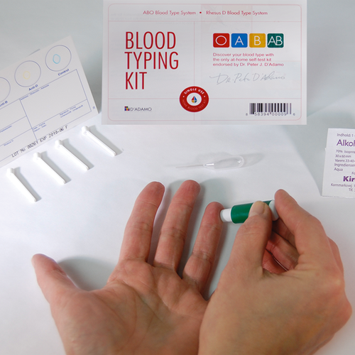
Affordable, FDA-approved kit that comes complete with everything you need to determine your blood type, including instructions, finger lancet and alcohol prep, plastic applicator sticks and testing card. Click to learn more |


The statements made on our websites have not been evaluated by the FDA (U.S. Food & Drug Administration).
Our products and services are not intended to diagnose, cure or prevent any disease. If a condition persists, please contact your physician.
Copyright © 1996-2024, Hoop-A-Joop, LLC, Inc. All Rights Reserved. Privacy Policy | Log In